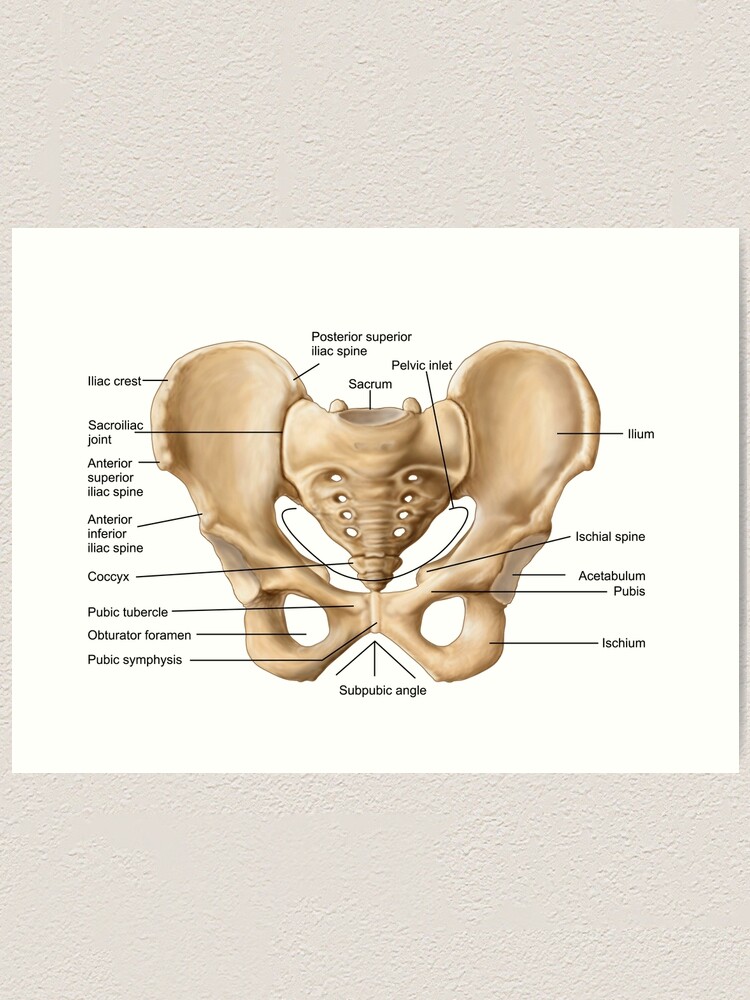
Pelvic Anatomy - Anatomy Of Pelvis - Human Anatomy Body. Anatomy of female pelvic area facebook twitter linkedin pinterest print fertility and reproductive health pelvic floor disorders fertility, pregnancy and childbirth women's health. It is strengthened and supported by several joints and ligaments. However, knowledge of the anatomy of various structures that surround these organs has evolved over time. The pelvis is the lower part of the torso. The pelvis is a basin shaped bony structure formed by the combination of two pelvic bones (hip bones or innominate bones) and the sacrum.
The male pelvic organs include the penis and various glands and ducts. The pelvis is the lower part of the torso. {{configctrl2.info.metadescription}} this site uses cookies. Laparoscopic anatomy of the female pelvic region. The pelvis's frame is made up of the bones of the pelvis, which connect the axial skeleton to the femurs, and therefore acts in weight bearing of the upper body.
PPT - Pelvic Anatomy PowerPoint Presentation, free ... from image5.slideserve.com Whenever someone talks about the pelvic floor in general, they are probably talking about these 5 muscles: Differentiate the different types of the female pelvis. Pelvic floor anatomy, pelvic floor therapy, physical therapist gift, pelvic floor specialist gift, physical therapy office art, pt art. The uterus represents the essential landmark of pelvic anatomy. The lining of the uterus. Describe the boundaries and subdivisions of the pelvis. The pelvis (plural pelves or pelvises) is either the lower part of the trunk of the human body between the abdomen and the thighs (sometimes also called pelvic region of the trunk) or the skeleton embedded in it (sometimes also called bony pelvis, or pelvic skeleton). The pelvic diaphragm is the third deepest layer of the pelvic floor which puts it at the very center of all the other muscles.
The male pelvic floor is a complex structure made up of muscles, ligaments, nerves and fascia.
The pelvis is inferior most part of the trunk. The pelvis is the lower portion of the trunk, located between the abdomen and the lower limbs. Describe the components & function of the pelvic diaphragm. Differentiate the different types of the female pelvis. This area provides support for the intestines and also contains the bladder and reproductive organs. This mri male pelvis axial cross sectional anatomy tool is absolutely free to use. The pelvis is a basin shaped bony structure formed by the combination of two pelvic bones (hip bones or innominate bones) and the sacrum. Ct body (lymph nodes) ct. Gross anatomy of the pelvis—namely the bladder, uterus, fallopian tubes, ovaries, rectum, and the muscles—has remained unchanged; The pelvis (plural pelves or pelvises) is either the lower part of the trunk of the human body between the abdomen and the thighs (sometimes also called pelvic region of the trunk) or the skeleton embedded in it (sometimes also called bony pelvis, or pelvic skeleton). The bony pelvis consists of the two hip bones (also known as innominate or pelvic bones), the sacrum and the coccyx. 5 out of 5 stars. It is strengthened and supported by several joints and ligaments.
The male pelvis is different from a female's. • divided into the true and false pelvis by the iliopectineal line. Describe the boundaries and subdivisions of the pelvis. 5 out of 5 stars. It is located in the middle of the pelvis between the urinary bladder lying before and the large bowel lying behind it.
Sherif Megahed - Pelvic Bone Anatomy from cdna.artstation.com The right and left hip bones, plus the sacrum and the coccyx, together form the pelvis. • pelvis begins at the iliac crests and ends at the symphysis pubis. The pelvis is inferior most part of the trunk. A pelvic ultrasound is a noninvasive diagnostic exam that produces images that are used to assess organs and structures within the female pelvis. The pelvis's frame is made up of the bones of the pelvis, which connect the axial skeleton to the femurs, and therefore acts in weight bearing of the upper body. Describe the boundaries and subdivisions of the pelvis. Whenever someone talks about the pelvic floor in general, they are probably talking about these 5 muscles: Gross anatomy of the pelvis—namely the bladder, uterus, fallopian tubes, ovaries, rectum, and the muscles—has remained unchanged;
On a sagittal plane, the uterus has a pyriform shape:
The right and left hip bones, plus the sacrum and the coccyx, together form the pelvis. During this time of information gathering about your hysterectomy options, there are a few terms that are helpful to know about pelvic anatomy. Using a speculum, a doctor can examine the vulva, vagina, and cervix.the strength of the pelvic muscles can also be tested. The pelvic diaphragm is the third deepest layer of the pelvic floor which puts it at the very center of all the other muscles. Describe the anatomy of the pelvic wall, bones, joints & muscles. Surgical anatomy of the female pelvis by laparoscopy. The pelvic bones are smaller and narrower. The pelvic girdle and pelvic spine. Describe the components & function of the pelvic diaphragm. Describe the boundaries and subdivisions of the pelvis. The male pelvic floor is a complex structure made up of muscles, ligaments, nerves and fascia. Consisting of the pelvic girdle and perineum, it supports the urinary and reproductive organs. {{configctrl2.info.metadescription}} this site uses cookies.
The uterus represents the essential landmark of pelvic anatomy. This mri male pelvis axial cross sectional anatomy tool is absolutely free to use. The lumbosacral plexus is formed by the lumbosacral trunk and the ventral rami of the first to third sacral nerves, and part of the fourth sacral nerve. Using a speculum, a doctor can examine the vulva, vagina, and cervix.the strength of the pelvic muscles can also be tested. Anatomy of female pelvic area facebook twitter linkedin pinterest print fertility and reproductive health pelvic floor disorders fertility, pregnancy and childbirth women's health.
"Anatomy of human pelvic bone." Art Print by ... from ih0.redbubble.net The lumbosacral plexus is formed by the lumbosacral trunk and the ventral rami of the first to third sacral nerves, and part of the fourth sacral nerve. The pelvic bones are smaller and narrower. The right and left hip bones, plus the sacrum and the coccyx, together form the pelvis. The pelvis is the lower portion of the trunk, located between the abdomen and the lower limbs. A pelvic ultrasound allows quick visualization of the female pelvic organs and structures including the uterus, cervix, vagina, fallopian tubes and ovaries. The nerves of the pelvis include: The pelvis is inferior most part of the trunk. The male pelvic organs include the penis and various glands and ducts.
Ultrasound uses a transducer that sends out.
The uterus represents the essential landmark of pelvic anatomy. The male pelvic organs include the penis and various glands and ducts. Laparoscopic anatomy of the female pelvic region. Describe the anatomy of the pelvic wall, bones, joints & muscles. By continuing to browse this site you are agreeing to our use of cookies. Pelvic floor anatomy, pelvic floor therapy, physical therapist gift, pelvic floor specialist gift, physical therapy office art, pt art. Consisting of the pelvic girdle and perineum, it supports the urinary and reproductive organs. The anatomy of the pelvis varies depending on whether you are male or female. 5 out of 5 stars. The bony pelvis consists of the two hip bones (also known as innominate or pelvic bones), the sacrum and the coccyx. Ct body (lymph nodes) ct. Ultrasound uses a transducer that sends out. It provides attachment to some important muscles in the region, and forms a cavity which accommodates several important internal organs.